Indian Scientists Develop Detailed Brainstem Atlas
Neuroscientific research has long been akin to early mapmaking, where scientists piece together a complex landscape from scattered observations. Despite advancements, much of the human brain's intricacies remain elusive, a challenge that pathologists face when diagnosing conditions like Alzheimer's disease. Typically, they examine limited samples from a brain comprising approximately 86 billion neurons.
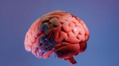
Researchers at the Sudha Gopalakrishnan Brain Centre (SGBC) at the Indian Institute of Technology, Madras (IIT-M) have made significant progress in this field. They have developed what is considered the most detailed three-dimensional atlas of the human brainstem at cellular resolution. This digital map, named Anchor (Atlas of Neurochemical Characterisation of the Human Brainstem with 3D Reconstruction), allows scientists to explore the brain seamlessly from MRI scans to individual nerve cells.
This atlas was constructed using over 500 tissue sections from foetal, childhood, and adult brains. Unlike costlier molecular methods, this tool relies on high-resolution microscopic images to detail the brainstem's structure, identifying more than 200 clusters of brain cells and nerve pathways. Eight chemical markers further differentiate cell types, offering a clearer view of this critical yet understudied brain region.
The Brainstem's Critical Role
The brainstem, though a small part of the brain, is vital for life. It connects the brain to the spinal cord and regulates essential functions such as breathing, heartbeat, sleep, and movement. Damage to even small clusters of cells in this area can have severe consequences, but its dense structure has historically made detailed mapping difficult.
Anchor's significance lies not only in its detailed anatomical mapping but also in bridging the gap between medical imaging and cellular pathology. Shubha Tole, a neuroscientist at the Tata Institute of Fundamental Research, describes it as an "unprecedented integration" of various scientific disciplines.
Traditionally, doctors examine the whole brain during autopsies or surgeries, starting with its folds and major structures. This method dates back to the pioneering work of Santiago Ramón y Cajal, over a century ago. While modern MRI scans provide views of the entire brain, they lack the cellular detail that microscopes can offer.
Advancements in Neurological Research
The Anchor atlas attempts to address this gap by allowing users to zoom from an MRI view of the brainstem down to individual neurons, maintaining precise spatial relationships. The atlas is freely accessible online, intended as a global reference tool for neuroscientists and neurologists. Its implications extend beyond anatomy, potentially aiding in understanding disorders like Parkinson's, Alzheimer's, and stroke.
According to Partha Mitra of Cold Spring Harbor Laboratory, detailed brain atlases could have a "transformative impact" on studying neurological diseases by showing how brains affected by conditions like autism differ from healthy ones. Additionally, the atlas might shed light on how infections, such as Covid-19, cause long-term neurological damage.
For instance, in the case of brain strokes, the atlas may reveal features that can help doctors preserve affected brain tissue, improving patient outcomes. Neurosurgeons could also use the atlas to navigate the brainstem more safely.
Creating a Comprehensive Brain Atlas
The development of this atlas is notable for its affordability and simplicity. It was created from high-resolution images of thin slices of post-mortem brain tissue, making detailed, cell-level mapping accessible. This effort was part of a broader shift in neuroscience, where progress increasingly relies on engineering and computational methods.
Over 20 scientists at SGBC spent 18 months analyzing more than 200 brain sections, combining MRI scans, microscopic anatomy, and 3D reconstruction into a single digital atlas. The SGBC now collaborates globally with over 200 researchers, engineers, and technicians.
While the human brain remains less thoroughly mapped than those of other species, the SGBC aims to image over 100 whole human brains across various life stages and neurological disorders. This initiative could create a reference library to show how diseases reshape the brain at a cellular level.
Though the new atlas won't solve the mysteries of the human brain, it offers researchers a more detailed map, enabling them to ask more informed questions.
Source: Original Article